Features
John Fulljames will join the University in November as Director of the Humanities Cultural Programme. We sat down with him to hear about his previous roles as Director of the Royal Danish Theatre and Opera and as the creator of a successful arts start-up; why he is so excited to work with the University and the City of Oxford; and his vision for the HCP.
Tell us about your experience as Director of the Royal Danish Theatre and Opera.
I have been in Denmark over the last five years, which has been an extraordinary time in the world – and in the world of culture. The pandemic has challenged the relationship between cultural institutions and audiences. But it has also offered enormous opportunity as societies have seen new value in coming together for shared live experiences. I have been really amazed and proud of the Royal Danish’s audience, which is stronger now than it has been for 15 years. That is truly remarkable given the time we have been through.
How did you achieve that?
I think the reason is that we were already very focused on growing a younger and more diverse audience by ensuring that the projects which we were putting on the stage would resonate with them and by building trust that we would deliver positive experiences. We had a strong focus on communicating clearly and transparently to a wide range of different audiences. I hope we can use a similar approach here to build audience trust in the programme of the Schwarzman Centre.
You also launched and ran a start-up company called The Opera Group. Tell us more about that?
The Opera Group was a company that I set up in my early 20s along with some amazing colleagues. It grew to become a regular commissioner of contemporary opera and a tourer in the UK and beyond and really flourished as an associate company at the Young Vic in London. I loved the experience of growing an organisation from scratch, and building the partnerships and networks necessary for it to thrive. We were so conscious of the value which the company gained from working in partnership with the established Young Vic, and also what the energy of our young company brought to them.
There sound to be parallels to where the Humanities Cultural Programme is in this early stage. What do you hope to apply from your previous experiences to the HCP?
The crucial thing about growing a new enterprise is to convene and nurture a brilliant team of people. We have the chance to do that here in Oxford. I see so much potential in building on the success of TORCH and the HCP over the last few years as we seek to establish the new venue with a rich and diverse programme. I see this as an opportunity not only to create something new for Oxford, but to work with a new generation of artists and companies and support them to do things which have never been done before!
What interested you most about this job?
It is unique to have a multi-disciplinary arts centre which will sit at the core of three different communities: the Oxford city and region, the university community, and the international cultural community. If we can grow the programme with all of these communities, so that they can all recognise themselves in it, we should also be able to offer each of them new opportunities to connect with each other. There is a fantastic cultural ecology in Oxford of which the new building and programme will become part. That’s a cultural ecology of individual artists as well as institutions such as theatres, music organisations and museums. I look forward to being part of that and hope to develop a range of local partnerships to complement the international partnerships we want to form.
How will you go about making your programming resonate with these audiences?
The Schwarzman will be a public building in the heart of the University. So we will start from the perspective of the audience – what do they want and what isn’t available in the local cultural ecology? We need to find ways of involving people not just as audiences but also as we incubate and develop projects here in Oxford. We can do this in a very local way – the Schwarzman Centre will be a building for Oxford with content developed in Oxford. At the same time, we can do it in an international way by presenting world-class content which would otherwise not be seen in the region and which can attract both live audiences and digital audiences from around the globe.
Talking of the building (which will open in 2025), what do you think of the plans for the performance venues?
The Schwarzman Centre will be the first purpose-built concert hall space on this scale in Oxford and we look forward to filling what promises to be a really world-class hall with a diverse range of music that brings in many different audiences. But this is not just a music venue, it is a genuine multidisciplinary building. It has been designed to be able to share theatre, dance, music, film and exhibitions. None of those art forms operate in isolation from the others and I think one of the benefits of the building will be the synergies which emerge from the enormous range of creativity going on inside. Part of the intention of the architecture is that value will be released from bringing the many different departments which make up the humanities together in a building which enables conversation between them. The same will be true for arts and performance.
What is the impact for a cultural institution of being part of a university like Oxford?
That is what will make the programme unique. The HCP’s identity flows from being at the heart of a centre of cutting edge of contemporary thinking. It offers us the richest possible context for exploring ideas and engaging people in ideas. The HCP has such an important role to play in helping to open up this historic centre of excellence so that its many riches can be visible to, valued by, and in turn enriched by, more people than ever before.
The Schwarzman Centre provides an opportunity for Oxford University to be porous in a new way, as a public building with a civic purpose. The HCP has to make sure that everyone can walk through the door and feel they are welcome and that they belong. I cannot wait to get started.
Whatever Hollywood might say, AI is not all about killer robots in a far off future. It is more mundane, more everyday - and much more ubiquitous. And thinking it just belongs in a sci-fi film is dangerous, since this leads to a sense it is not relevant or that it is even unreal. Professor Ursula Martin bristles slightly at the very idea of robots, killer or otherwise, and points out that everyone’s lives are already affected by AI – and there are ‘bigger things at play’ than people realise.
The Maths professor and computer expert has put together a fascinating exhibition in the Bodleian about the history of AI – and has raided Oxford’s collections for treasures which showcase thinking about AI, illustrate fundamental ideas, and provoke debate.
AI is very useful. It’s all around us...It is changing our lives...Making...AI all about killer robots gets us off the hook of taking responsibility and distracts attention from the more everyday reality of AI
Professor Ursula Martin
Victor Frankenstein played God, she says, and Mary Shelley’s manuscript still has the power to shock. In the exhibition, it is open at the page where 'by the glimmer of the half-extinguished light' Frankenstein sees for the first time 'the dull yellow eye' of the creature he has created in his laboratory. Another exhibit is Ada Lovelace, describing Charles Babbage’s calculating machines. She places them in a contemporary theological debate: did the the creator of the machines challenge God, or merely help us to understand His works better?
Meanwhile, Ramon Lull’s colourful medieval diagrams present simple reasoning as an almost mystical process. But, for the 19th century economist Stanley Jevons, the goal was more mundane. His 'Logic Piano', a construction of ivory, wood, wire, showed, in principle, it was possible to mechanise human reason itself.
 The logic piano
Jonathan Swift, meanwhile, imagined a machine that could write a book on any topic ‘without the least assistance from genius or study’. And, in the early days of the computer, Christopher Strachey experimented with simple computer generated love-poems: the fore-runner of today’s AI software. It can mine millions of existing texts and 'learn' rules to create 'plausible' new texts about any given topic. Strachey’s programme used a very restricted vocabulary, which gave the poems an oddly prim and stilted tone.
Similarly, it is all too easy for modern AI to reflect biases in the texts it has learned from, propagating and amplifying those biases.
Professor Martin points out, to deliver modern AI involves vast quantities of data, and fast computers that use clever algorithms, not just for calculation, but to reason and find patterns too. Looking at early examples, such as Strachey’s poems, shows just how straightforward some of the underlying ideas are. According to Wadham College-based Professor Martin, it is the scale of the data and the power of the computation that transforms these simple ideas into present-day AI. Understanding them in context can give us new ways to think about contemporary concerns as well.
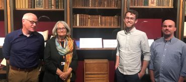 Professor Martin and the team
‘There are so many stories you can tell from the Bodleian archives,’ Professor Martin says enthusiastically. ‘We’re trying to tell the history of AI here in 15 objects.'
So it is emphatically not all killer robots. Professor Martin adds, ‘AI is very useful. It’s all around us, for example your phone or your satnav or your bank are full of AI. It is changing our lives, and both as individuals and as society as a whole we need to think and act responsibly, just as we should with any other technology. Making conversations about AI all about killer robots gets us off the hook of taking responsibility and distracts attention from the more everyday reality of AI.’
Dr Qiang Zhang of the Radcliffe Department of Medicine explains how artificial intelligence is being used to help researchers and physicians interpret medical imaging.
Disruptive AI-based imaging technology might replace the injection of dye ‘contrast agents’ usually needed to show clear images of scar of the heart
Imagine you are a medical doctor, faced with a patient with suspected heart disease for symptoms such as chest pain, tightness, or shortness of breath. One way to find out what is happening, and help guide patient prognosis, is to do a cardiovascular MRI scan to look into any heart muscle abnormalities. The scan involves injecting a ‘contrast agent’ (a dye that will improve image contrast and show up scars on images) into a vein in the patient. Contrast-enhanced MRI has been the clinical standard to provide clear scar images, but it’s painful, and makes already expensive MRI scans even more so.
What’s more, this method is limited in patients with significant kidney failure – their kidneys have difficulty clearing the dye from their bodies, sometimes leading to irreversible complications. Some patients will be allergic to the contrast agent, and you might want to limit the use of injectable contrasts in some patients, such as pregnant women and children.
So how do you find out about what might be going on in your patient’s heart in that case, without injecting into them a contrast agent?
It turns out that injecting a contrast agent might not be the only way to get clear MR images to reveal scars in the heart muscle – in 2010, Professor Stefan Piechnik from the Radcliffe Department of Medicine at Oxford University came up with a method to study heart muscle properties, using a contrast-free MRI technique called T1-mapping. It produces an image of the heart with numerical values that change with different diseases.
Such contrast-free MRI contain a lot of information about heart tissue properties, some of which is subtle, or difficult to identify as a scar or other pathologies. As of now, researchers are still exploring the best ways to interpret and display the information from these contrast-free T1-maps, which is one of the reasons that they are not yet widely used by medical doctors.
This is why our cross-disciplinary team of AI scientists, magnetic resonance imaging specialists and cardiologists at the University of Oxford worked to find ways that artificial intelligence (AI) can enhance these contrast-free MRIs, to produce clear images of heart muscle scarring. AI effectively works like “virtual contrasts” to replace conventional intravenous contrasts.
We developed an AI-powered algorithm to combine multiple contrast-free MR images together with heart motion information, enhance the pathological signals in them, to reveal scars in a similar way to conventional contrast-enhanced MRI. This technology is called “virtual native enhancement”, or VNE, as it acts as an enhancer for the MR images, using only the ‘native’ (ie, non contrast agent enhanced) images produced by an MRI scanner.
In 2021, our team released the first proof of concept for this idea, by detecting scars in the heart muscle for patients with hypertrophic cardiomyopathy, a common genetic heart disease affecting 1 in 500 people, and the most common cause of cardiac death among young people.
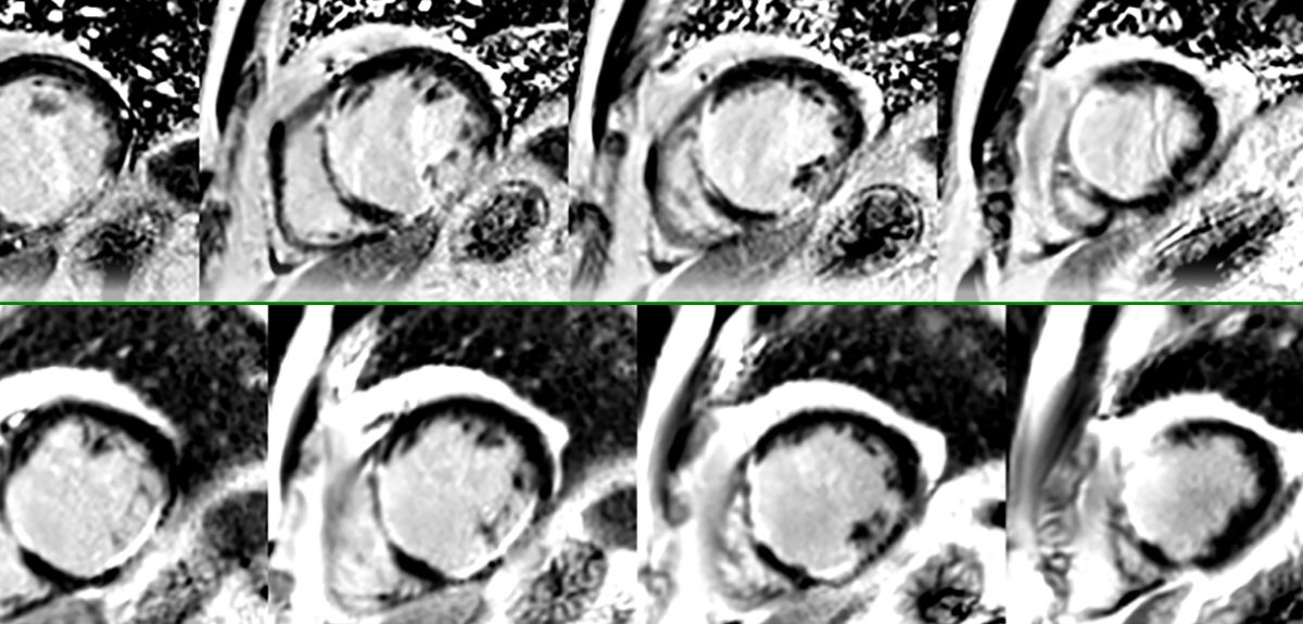
Recently, we have found that VNE can also detect scars in patients who have had a heart attack. We compared contrast-free VNE with conventional contrast-enhanced MRI in these patients. We found that VNE highly agreed with the conventional MRI in detecting previous heart attack scars and their extent. Additionally, the VNE image quality was actually better, all without the patients needing to receive an injection.
Once completely validated, this new technology may slash the time that patients need to spend in an MRI scanner from the standard 30-45 minutes to within 15 minutes, saving more than half the scan cost, yet producing images that are clearer, more diagnostically useful, and easier to interpret.
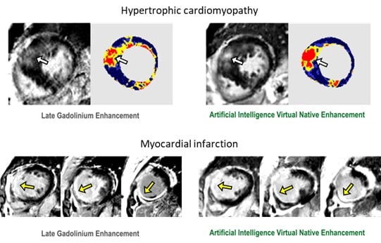
Image: Development of VNE in detecting heart muscle scars for two different heart diseases. The right panels show our new contrast-free method, while the left panels show conventional contrast-enhanced which requires injecting contrast agents. Arrows point to the detected scars.
We think that these successive breakthroughs mark the beginning of a new era of diagnostic medical imaging, using AI instead of IV contrasts to reveal pathologies in the human body: we might finally be able to get rid of injections when it comes to heart MR imaging.
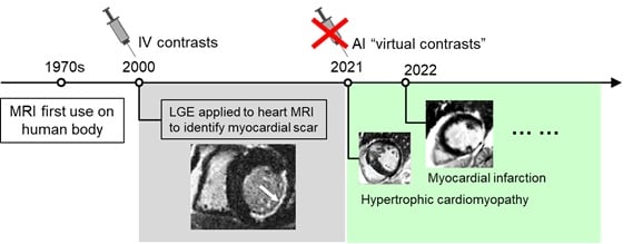
Image: Background of IV contrasts of MRI, and the emerging new era of AI “virtual contrasts”.
We are now working to further improve the capabilities of this technology, to detect more complex heart diseases and their underlying mechanisms, beyond the diagnostic power of current MRI. We plan to use these methods in large clinical studies as a diagnostic tool for novel investigations.
We think that this kind of Virtual Native Enhancement technology is an exciting and potentially game-changing advance for clinical MRIs in the future. Patients going in for a clinical MRI scan might not need an injection for most MRI scans, not just for the heart, but potentially for other organs as well. This would cut costs for healthcare providers, meaning that many more patients could access MRI scans; the risks of contrast-agent injections complications would disappear too. We hope adoption of this method could contribute to the digitalization of the NHS, something which is very much needed to address the backlog post COVID-19 pandemic.
With thanks to Professor Stefan Piechnik and Professor Vanessa Ferreira, joint senior authors of the study.
Further reading in Circulation: Zhang et al 2021, Zhang and Burrage et al 2022.
Alberto Lazari of the Nuffield Department of Clinical Neurosciences explains the importance of insulation in our brains' wiring.
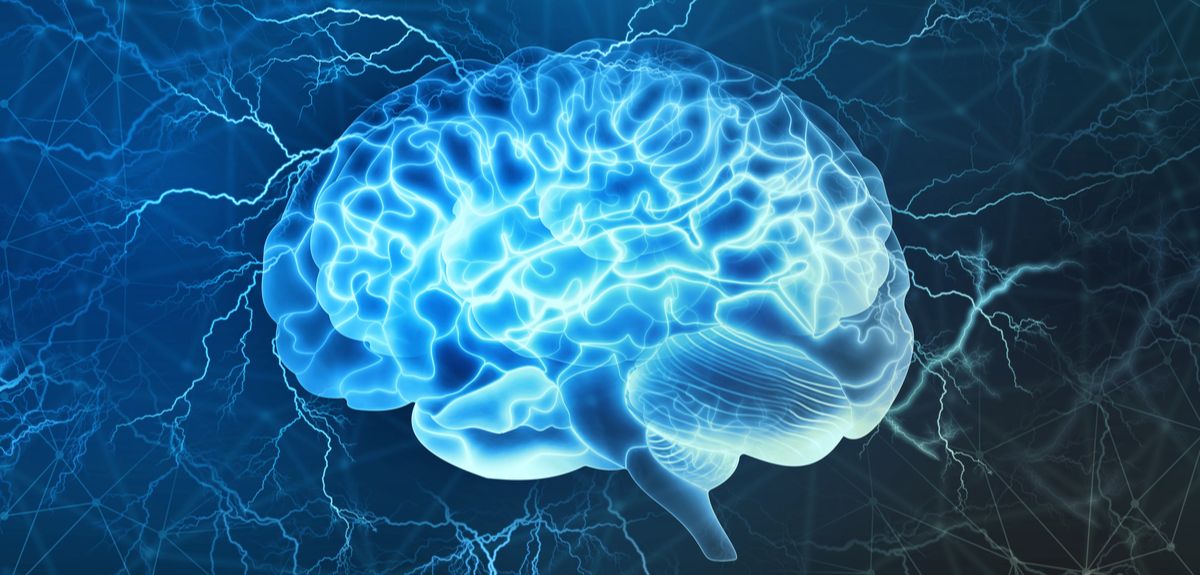
Our brains contain a striking amount of ‘brain wires’, which allow electrical signals to send important information from one corner of the brain to another. Although these brain wires are made up of biological material, they also bear surprising resemblances to the electrical wires you can see when you do a DIY job in your home. For instance, one key feature that allows the brain wires to work is that they are tightly insulated. A little bit like metal wires are coated with plastic, brain wires are also wrapped in an insulation material, called ‘myelin’. Myelin is essentially a fatty layer of insulation, wrapped around many of the wires in your brain.
Myelin is incredibly important. When this insulation layer breaks down, the brain struggles to transmit signals at its usual speed, which is what happens in conditions like multiple sclerosis. However, the insulation of brain wiring has often been overlooked by scientists. It is particularly difficult to measure non-invasively in a live human. On top of that, this insulation has long been considered a static part of the brain which is not particularly relevant to understanding the brains of healthy adults. While myelin is clearly important in multiple sclerosis, until recently very few scientists had studied myelin beyond the realm of disease.
However, recent studies have now called some assumptions about myelin into question. In particular, in the past decade many labs around the world, including here at Oxford, have shown that myelin is more complex and dynamic than previously thought. Ground-breaking new methods have also been developed to effectively measure fat-rich insulation through magnetic resonance imaging (MRI), allowing us to ask new questions about this ever-elusive insulation layer that envelops our brain wiring.
For example, we know that everyone has a different brain, and brain wiring is one way that our brains differ from each other. Do different people have different levels of wiring insulation? And do these differences between individuals influence how our brains work? As simple as these questions may sound, they had not been asked before - until now.
At the Wellcome Centre for Integrative Neuroimaging, we set off to find an answer, using new MRI techniques to study myelin. First, we scanned a large group of participants and captured detailed MRI brain scans which gave us information about myelin. We then tested the same participants with a type of non-invasive brain stimulation called Transcranial Magnetic Stimulation, or TMS. Using TMS, we can create fast electrical signals and track them across the brain on a millisecond scale. This technique allowed us to capture fast electrical communications between brain areas – even those on opposite sides of the brain. This was particularly useful, because this very rapid electrical communication along brain wires is exactly what we expect would be influenced by insulation, very much in the way that the insulation of metal wires in our homes changes their electrical conductance.
Our findings showed for the first time that variation in brain wiring insulation between people is associated with significant differences in how brain areas communicate. For example, participants with more myelin in a given “brain wire” connecting two brain regions also tend to have a stronger electrical connection between those two brain regions. This is important because it confirms the significance of myelin not just to disease, but also to the everyday functioning of the brain. It also demonstrates the utility of studying myelin to understand the fine details of how different regions of the human brain communicate with each other.
Finally, our results also carry important practical implications. If our individual brain wiring insulation is linked with how we respond to brain stimulation, could information about myelin be used in the future to study clinical responses to brain stimulation? For example, TMS is already being used as a promising therapy for major depression, but with huge variability in how people respond to this treatment. Could information about brain wiring insulation tell us more about why some people respond better than others to TMS? And could this eventually help us better tailor treatment? We still do not have answers to these questions. However, what is certain is that this fat-rich insulation of our brain wiring, once thought to be a totally uninteresting part of the brain, is likely to have some more exciting surprises in store for us.
The paper, 'A macroscopic link between interhemispheric tract myelination and cortico-cortical interactions during action reprogramming', can be read in Nature Communications.
Professor Chrystalina Antoniades of the Nuffield Department of Clinical Neurosciences explains how the COVID pandemic accelerated an innovation in one research project into Parkinson's Disease.
Parkinson’s is a progressive neurological condition, which affects around 145,000 people in the UK.
Symptoms start to appear when there isn’t enough of the chemical dopamine in the brain to control movement properly. People with Parkinson’s don’t have enough dopamine because some of the nerve cells that make it have died.
There are lots of symptoms, but the three main ones are tremor (shaking), slowness of movement, and rigidity (muscle stiffness).
Doctors typically diagnose and monitor the progression of Parkinson’s by assessing these symptoms using a ‘clinical rating scale’. This relies solely on the clinician’s own subjective impression of the person’s condition.
Since 2016, the NeuroMetrology Lab at the University of Oxford has been developing objective numerical measures to help doctors accurately diagnose disease and monitor the progression of Parkinson’s – which could lead to the provision of more targeted and timely treatment. Until recently this research team, based in the Nuffield Department of Clinical Neurosciences, has been carrying out their research via in-person clinics, attended by patients four times a year.
During the patient’s two-hour clinic visit, the researchers would measure subtle abnormalities in the speed and coordination of fast eye movements (known as saccades), hand movements, and gait. They would also assess cognitive performance using tasks on a tablet. Then they would try to work out whether these numerical measures could accurately and objectively quantify Parkinson’s, and track its progression over time.
The advent of home monitoring
One of the features of Parkinson’s symptoms is that they fluctuate both throughout the day and from day to day. So the research team always knew that they wanted to be able to monitor symptoms at home as well as in the clinic. They were aware that patients’ behaviour during short clinic visits every few months was probably not representative of the condition’s progression overall.
In 2020, the Covid pandemic put an immediate stop to research with human participants, making in-person clinics impossible. This apparent disaster in fact accelerated the researchers’ plans to roll out wearable technology and enable study participants to monitor their symptoms at home.
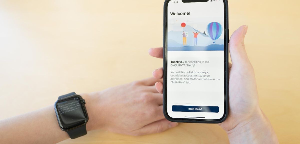
The biopharmaceutical company MSD is funding this new phase of the research project. The new grant has enabled the team, which I am leading, to work with MSD and the technology company Clinical Ink to capture data on participants’ symptoms at home. The wearable technology combines an Apple watch and phone to test a range of both motor and cognitive aspects of Parkinson’s.
I am delighted to be able to offer to our research patients the opportunity to be monitored so closely by such clever technology. My team has been working hard to make this a pleasant experience for all our patients and we are incredibly honoured to have such tremendous support from the Parkinson’s Disease community.
‘Digital health technologies offer tremendous opportunity to measure and objectively quantify the symptoms and progression of neurological disease,’ said Dr Marissa Dockendorf, Executive Director, Head of Global Digital Analytics and Technologies at MSD Research Laboratories. ‘MSD is excited to collaborate with the University of Oxford to further the development and characterisation of digital measures to support timely and reliable evaluation of potential new treatments for Parkinson's disease.’
How patients can monitor their symptoms at home
A member of the team sets everything up with participants remotely during a telemedicine appointment, explaining how to use the watch and the app on the phone. The participants receive instructions and the app gives step by step guidance on what to do. Participants are required to carry out testing at home once a month, performing tasks on the app such as reading, testing reaction times, and cognitive tasks.
David Williams, a participant in the study, said: ‘The wearable technology is very easy and comfortable to use. The instructions are very clear, the exercises are well explained and not at all difficult to accomplish. The staff are friendly, approachable people who always leave me with a sense of being valued as a contributor to what is obviously a very important research study. If you’re at all anxious about taking part, don’t be, just sign up. You won’t regret it!’
Kevin McFarthing, another participant, also stressed how easy it was to carry out home monitoring: ‘The OxQUIP team is very professional and thoroughly well organised, and are a pleasure to work with. They did a great job training me to use the remote devices’, he said.
The home monitoring does not replace clinic visits entirely; patients have a telemedicine appointment every four months, as well as the opportunity to come in to an in-person clinic if they wish.
Joan Severson, Chief Innovation Officer at Clinical Ink, said: ‘We are honored that our mobile and wearable technology plays an integral role in this study of Parkinson’s disease in Oxford. We are excited to collaborate with researchers who tirelessly work to increase objective numerical measures for diagnosing and monitoring disease progression.’
Looking to the future
The Covid pandemic was a dark period for many, and yet it accelerated this change in the way this research project is being carried out. The team is now able to gather richer, more nuanced and accurate data to feed into their analysis.
The outcomes of this project will improve the diagnosis, tracking and treatment of Parkinson’s. The insights gained about monitoring disease progression will make the assessment of clinical trials more efficient, leading to faster drug discovery not only for Parkinson’s, but potentially for a range of neurological conditions.
This work is part of the OxQUIP (Oxford Quantification in Parkinsonism) programme. If you’re interested in taking part in this study, please email [email protected].
- ‹ previous
- 9 of 248
- next ›

 Teaching the World’s Future Leaders
  A blueprint for sustainability: Building new circular battery economies to power the future
 Oxford citizen science project helps improve detection of antibiotic resistance
 The Oxford students at the forefront of the fight against microbial resistance
  The hidden cost of AI: In conversation with Professor Mark Graham
  Astrophoria Foundation Year: Dr Jo Begbie reflects on the programme’s first year
 World Malaria Day 2024: an interview with Professor Philippe Guerin
 From health policies to clinical practice, research on mental and brain health influences many areas of public life
 From research to action: How the Young Lives project is helping to protect girls from child marriage